A multi-site experiment
This work was carried out with the MACH3CANCER consortium. The aim was to understand the different ways cancer cells organise and behave in complex 3D environments, and how they respond to treatments.
It is a rare thing for the same experiment to be performed independently at different research institutes within a single study. That was a defining feature of this project. We wanted to understand how much experiments could vary between laboratories while still producing consistent results.
As a way to study this, we modified a cell line originally created by Malin Pedersen, where NRAS activation drives MAP kinase signalling.[1] The cells grow well as 3D spheroids, and we added an ERK sensor so we could measure signalling responses to treatment throughout them. The ERK sensor came from a period of development in fluorescent signalling reporters, when researchers were finding new ways to turn signalling activity into visible changes inside living cells.[2]
The NRAS activation increases the activity of the MAP kinase pathway, a signalling cascade from RAS or RAF, through MEK, to ERK. This pathway controls processes including cell proliferation. In 2002, Mike Stratton and colleagues at the Wellcome Trust Sanger Institute discovered that BRAF is frequently mutated in melanoma, in work that also involved researchers at the Institute of Cancer Research.[3] This helped lead to treatments that target MAP kinase signalling in melanoma. These MAP kinase pathway-targeted treatments include binimetinib, the MEK inhibitor we used in our study.

A new kind of 3D imaging
We started developing the protocol for these experiments around 2022 using dOPM, developed in the Photonics Group at Imperial.[4] We worked out the practicalities of imaging and adding treatments, and developed a protocol that researchers at four centres in the UK and Spain could use independently.
We used dual-view oblique plane microscopy (dOPM), a form of light-sheet microscopy designed for rapid volumetric imaging using a single objective, to image the melanoma spheroids in 3D.[4]
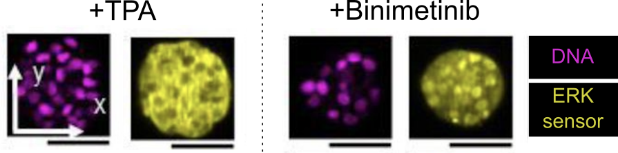
Our ERK sensor changes its location within the cell. When ERK signalling is turned down, the sensor moves into the nucleus, allowing us to measure signalling throughout the 3D spheroids.
Reproducible across laboratories
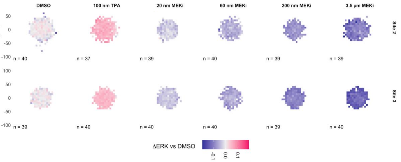
The protocol turned out to be robust. The major treatment responses were reproducible across sites and remained consistent across experimental differences such as laser intensity. I hope we can expand on this approach with models of a wider range of cancers, more targeted therapies and therapeutic combinations, and extended live imaging to see how responses to treatment progress over hours and days of treatment.
Groups involved
We are from different research groups across four experimental centres, together with the team that developed the dOPM technology:
- Tumour Cell Biology Laboratory — Erik Sahai · Francis Crick Institute
- Edinburgh Drug Discovery — Neil Carragher · Institute of Genetics and Cancer, University of Edinburgh
- Dynamical Cell Systems — Chris Bakal · Institute of Cancer Research
- Colorectal Cancer Laboratory — Eduard Batlle · IRB Barcelona
- Biophotonics Group — Imperial College London · dOPM development
Sharing the data
The raw data we generated is openly shared. This kind of 3D imaging data is still relatively rare, and it can be used by others to train image analysis algorithms, compare with other datasets, or reuse to ask new questions.
You can explore and download the dataset from the BioImage Archive.
References
- Pedersen M, et al. Primary melanoma of the CNS in children is driven by congenital expression of oncogenic NRAS in melanocytes. Cancer Discovery (2013). doi:10.1158/2159-8290.CD-12-0464
- Regot S, et al. High-sensitivity measurements of multiple kinase activities in live single cells. Cell (2014). doi:10.1016/j.cell.2014.04.039 · ERK-KTR-mRuby2 on Addgene
- Davies H, et al. Mutations of the BRAF gene in human cancer. Nature (2002). doi:10.1038/nature00766
- Sparks H, Dent L, Bakal C, Behrens A, Salbreux G, Dunsby C. Dual-view oblique plane microscopy (dOPM). Biomedical Optics Express (2020). doi:10.1364/BOE.409781
You can read our report about the study here.